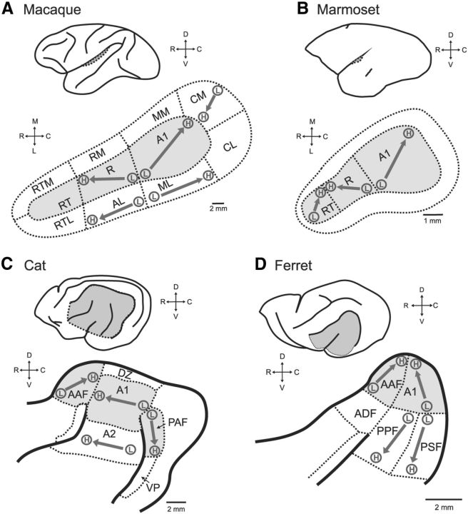
Figure 1.
Schematics of auditory cortex across four species: A, Macaque; B, marmoset; C, cat; and D, ferret. In each panel, the top schematic shows an outline of the brain with auditory cortex indicted (dotted line, gray). The bottom schematic in each panel shows a closer view of the auditory cortex, with sulci (solid lines) and field boundaries (dotted lines). Core auditory cortex is shaded gray, and the orientations of known tonotopic maps are indicated with an arrow from low (L) to high (H) frequencies. A1, Primary auditory cortex; A2, secondary auditory area; AAF, anterior auditory field; ADF, anterior dorsal field; AL, anterolateral belt; CL, caudolateral belt; CM, caudomedial belt; DZ, dorsal zone; ML, mediolateral belt; MM, mediomedial belt; PAF, posterior auditory field; PPF, posterior pseudosylvian field; PSF, posterior suprasylvian field; r, rostral field; RM, rostromedial belt; RT, rostral temporal field; RTL, rostrotemporal lateral belt; RTM, rostrotemporal medial belt; VP, ventral posterior field.

