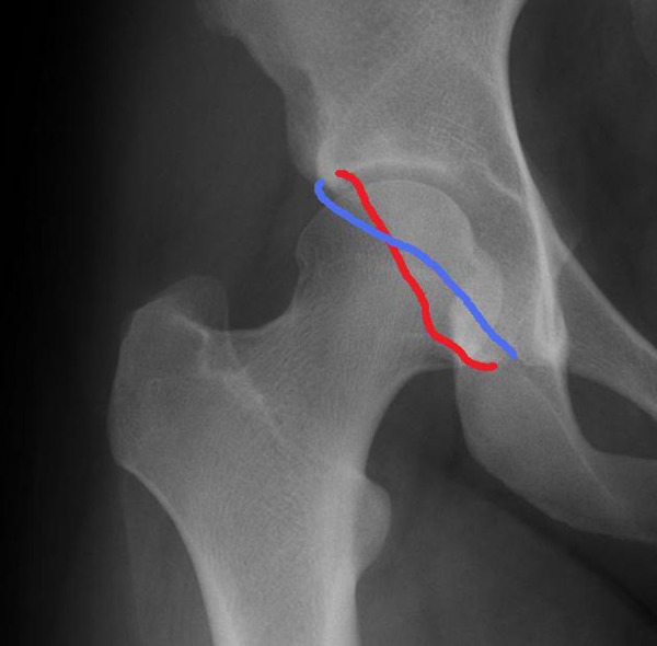
Figure 3.

“Crossover” sign: Anteroposterior radiographs were used to determine the presence of a crossover sign, which is consistent with a pincer lesion. The anterior wall is outlined in red and the posterior acetabular wall in blue. Typically, the anterior wall remains medial to the posterior wall. If the anterior wall crosses the posterior wall and becomes more lateral than the posterior, this is considered a crossover sign.
