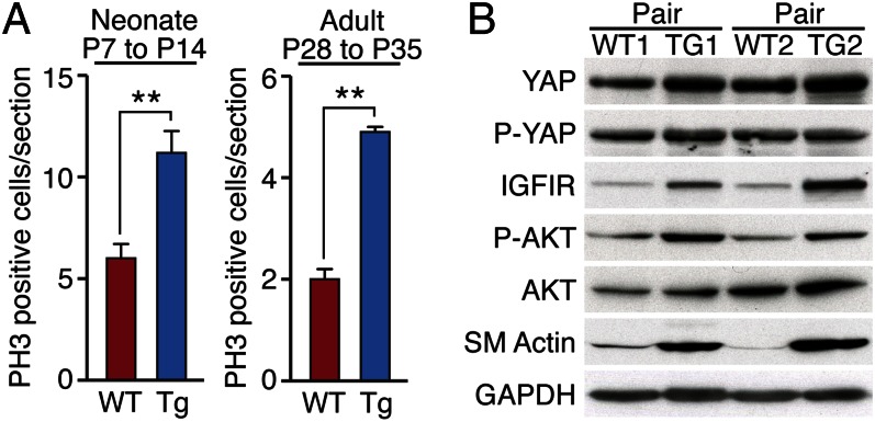
Fig. 6.
Yap promotes cardiomyocyte proliferation after MI. (A) Quantification of PH3 immunostaining of WT and αMHC-YapS112A Tg hearts at 7 d post-MI performed on P7 mice (neonates) or at 7 d post-MI performed on P28 mice (adults). Three hearts were analyzed for each genotype at each time point. Data are mean ± SD. **P < 0.005. (B) Western blot analysis comparing the indicated protein levels in two pairs of WT and αMHC-YapS112A Tg adult hearts. GAPDH was detected as a loading control.

