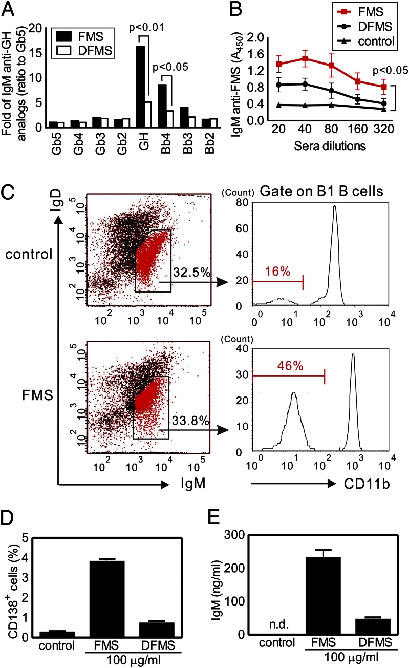
Fig. 4.
Correlation between anti-glycan IgM production and B1 B cells expansion in the mice immunization with our Reishi polysaccharides. (A and B) Antisera from FMS- and DFMS-treated mice were assessed by Globo H-related printed glycan microarray (A) and FMS-coated ELISA plate (B). (A) Binding of IgM to Globo H (GH) and its truncated forms (tested at 1:100 dilution) was normalized by setting the IgM anti-Gb5 as 1. (B) Binding of IgM to FMS (tested at 1:20–1:320 dilution) was measured by detecting the absorbance at 450 nm. (C–E) Expansion of peritoneal B1 B cells upon FMS immunization. FACS profiles of B1 B cells represent FMS-treated mice and control. Additional levels of B2 B cell and macrophage are shown in Fig. S4. Numbers (%) indicate the positive cells in each gate (C). FMS induced up-regulation of plasma cell surface marker (CD138) (D) and IgM production (E) in ex vivo B1 B cells culture purified from FMS-treated mice. Means ± SD (n ≈ 3–5 for each experiment). n.d., not detectable.

