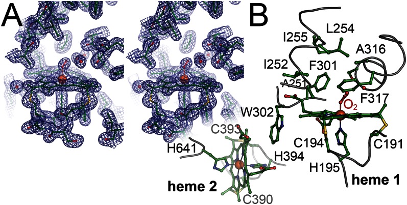
Fig. 3.
Heme group environment in RoxA. (A) Stereo representation of the distal pocket at heme group 1 with a bound O2 molecule. The displayed 2Fo – Fc electron density map is contoured at the 1 σ level. (B) Amino acid residues at and around the two heme groups. Three regions of the protein form a spacious, hydrophobic cavity above the distal side of heme 1. W302, a residue also conserved in CcpA peroxidases, bridges the hemes. Heme group 1 is linked to the protein via C191 and C194, with H195 as a proximal axial ligand, and heme group 2 is attached through C390 and C393, with H394 as a proximal axial ligand and H641 as a distal axial ligand.

