Fig. 4.
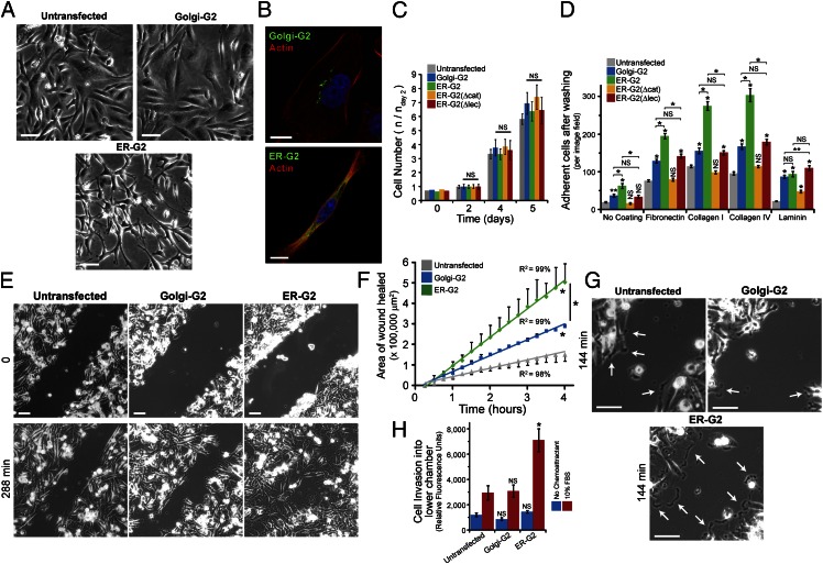
ER-targeted GalNAc-T2 enhances cell adhesion and migration. (A) Phase-contrast images of untransfected and transgenic GalNAc-T2 cells. (Scale bars, 100 µm.) (B) Actin staining in Golgi-G2 and ER-G2 cells. (Scale bars, 10 µm.) (C) Mean growth rate ± SEM of model GalNAc-T2 cells. (D) Mean adhesive strength ± SEM of model GalNAc-T2 cells. *P < 0.01, **P < 0.05 relative to untransfected or stated samples. (E) Migration assay using scratch wound of cellular monolayer. (Scale bars, 100 µm.) (F) Mean of wound closure ± SEM (n = 2 experiments). *P < 0.01 relative to untransfected cells or stated samples. (G) Lamellipodia (arrows) in migrating ER-G2 compared with untransfected and Golgi-G2 cells. (Scale bars, 100 μm.) (H) Transwell migration assay in a Boyden chamber (n = 3 experiments). *P < 0.01 relative to untransfected cells. Nuclei stained using Hoechst.

