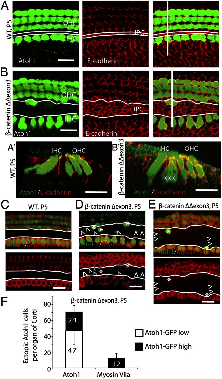
Fig. 3.
Hair cells differentiate from cells that express high levels of Atoh1. (A) Atoh1 was expressed in hair cells, and E-cadherin was expressed in supporting cell junctions in a wild-type organ of Corti. The white lines along the longitudinal axis of the organ of Corti indicate inner pillar cells. (A′) An x–z scan of A was performed at the white line perpendicular to the longitudinal axis. (B) In β-catenin–overexpressing littermates the number of pillar cells increased (between the white lines), and Atoh1-positive cells appeared in the pillar cell region at P5 in mice administered tamoxifen at P1. (B′) An x–z scan of B (at the perpendicular white line). (C) Atoh1 and myosin VIIa were expressed in hair cells in a wild-type organ of Corti at P5. No Atoh1- or myosin VIIa-positive cells were seen in the pillar cell region (white outlines). (D) β-Catenin expression induced ectopic Atoh1-positive cells in the pillar cell region, some of which showed high levels of Atoh1 (asterisks) and expressed myosin VIIa. Cells with low levels of Atoh1 (arrowheads) did not express myosin VIIa. (E) In β-cateninΔΔexon3 mutants, some of the ectopic cells that expressed high levels of Atoh1 in the expanded pillar cell region expressed myosin VIIa. (F) Quantification of ectopic Atoh1- and myosin VIIa-positive cells in the pillar cell region in β-cateninΔexon3 mutants. One-third of Atoh1-positive cells showed high-level expression of Atoh1 (black bar) (n = 3). (Scale bar, 20 μm.)

