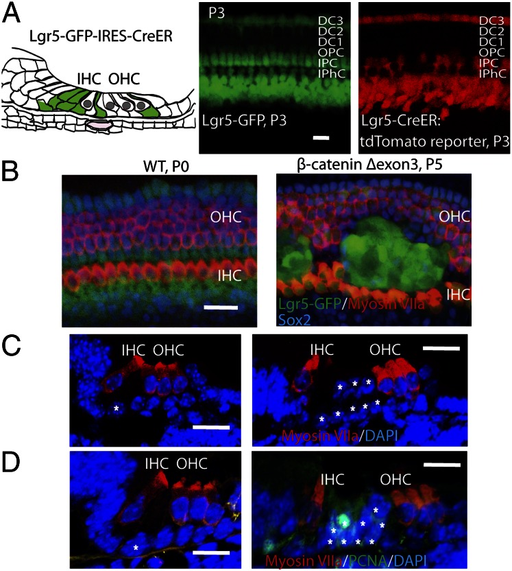
Fig. 4.
Wnt signaling expanded Lgr5-expressing supporting cells in vivo. (A) Lgr5 is expressed in a subset of supporting cells as illustrated. Third Deiters’ cells, inner pillar cells, and cells in the greater epithelial ridge expressed GFP in the Lgr5-GFP-IRES-Cre-ER mouse. Cre activity was seen in Lgr5-expressing cells at P3 when the mice were crossed with a Cre-activated tdTomato reporter mouse after tamoxifen administration at P1. (B) Expansion of Lgr5-positive cells at P0 upon activation of β-catenin signaling at E16.5 in Lgr5-Cre-ER mice crossed to β-cateninflox(exon3) mice, compared with wild-type mice. (C) The pillar cells were expanded in the organ of Corti cross-section. (D) The cells expanded in the pillar cell area were positive for PCNA (white asterisks indicate pillar cells). (Scale bar, 20 μm.)

