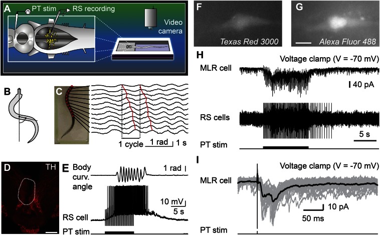
Fig. 3.
Stimulation of the PT activates brainstem locomotor circuits. (A–E) In a semi-intact preparation, the PT was stimulated and the reticulospinal (RS) cells were recorded together with locomotor movements. (B and C) Body curvature oscillations during locomotion. (C) PT stimulation (10-s train, 5 Hz, 11 µA, 2-ms pulses) elicited swimming as illustrated by the rostrocaudal mechanical wave. (D) The PT stimulation site was confirmed histologically by an electrolytic lesion (enclosed by white dashed line) that colocalized with TH-immunoreactive neurons (red). (Scale bar: 200 µm.) (E) PT stimulation elicited reticulospinal activity together with swimming. Stimulation artifacts were clipped. (F–I) Whole-cell patch-clamp recordings of MLR neurons retrogradely labeled from an injection of dextran amines conjugated to Texas red (MW, 3,000 Da) in the MRRN. (F and G) MLR neuron labeled by the Texas red–dextran amine injection in the MRRN and by the fluorescent marker added to the patch pipette solution (Alexa Fluor 488 hydrazide). (Scale bar: 10 µm.) (H) Trains of stimuli (10-s train, 5 Hz, 7 µA, 2-ms pulses) activated the whole-cell patch-clamped MLR neuron concomitantly with reticulospinal neurons, which were used to monitor locomotor activation. Action potentials were recorded extracellularly from reticulospinal neurons of the MRRN, and stimulation artifacts were clipped. (I) Single pulse PT stimulation (0.1 Hz, 7 µA, 2-ms pulses) elicited short-latency excitatory postsynaptic currents in MLR neurons. (H and I) Data from two different animals.

