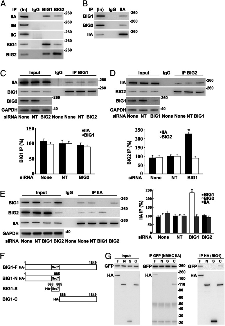
Fig. 1.
Co-IP of endogenous NMHC IIA with BIG1 and BIG2: co-IP of BIG2 with IIA increased after BIG1 depletion. Samples [5%, Input (In)] of HeLa cell proteins used for IP (100 µg) and immunoprecipitated proteins were analyzed by Western blotting with indicated antibodies and densitometric quantification. (A) Proteins from IP with control IgG (50%) or antibodies against BIG1 (25%) or BIG2 (50%) reacted with antibodies against NMHC IIA, -B, or -C or BIG1 or -2. (B) Proteins (50%) from IP with NMHC IIA (IIA) antibodies or control IgG reacted with BIG1 or -2 or IIA antibodies. (C) Effects of BIG2 siRNA depletion on IP of BIG1 (25%) or with control IgG (50%). (D) Effects of BIG1 depletion on IP with control IgG or BIG2 antibodies (50%). Data are reported as in C. *P < 0.005 vs. NT. (E) Effects of BIG1 or BIG2 depletion on IP of IIA (25%) or with control IgG (50%). Data are reported as in C, with representative blot on left. *P < 0.01 vs. NT. (F) Diagram of N-terminal-tagged full-length BIG1 and fragment sequences used in plasmids (pCMV-HA vector) plus EGFP-NMHC IIA to cotransfect cells 24 h before preparation of extracts. (G) Samples of extracts before IP (Input) and of proteins from IP (25%) with antibodies against GFP (IIA) or HA (BIG1) were reacted with tag antibodies on Western blots.

