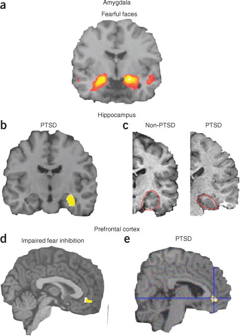
Figure 4.

Human neural circuitry involved in fear-related disorders and PTSD. (a) Viewing of fearful or angry faces (compared to shapes) robustly activates human amygdala across protocols and cohorts (reproduced with permission from ref. 27). Often this amygdala activation is increased in fear-related disorders. (b) Right hippocampal activity is lower in youths with post-traumatic stress symptoms than in healthy controls (reproduced with permission from ref. 38). (c) Reduced hippocampal volume in a patient with PTSD (right) compared to that in a subject without PTSD (left). Hippocampus outlined in red (adapted with permission from ref. 39). (d) Reduced neural activation of vmPFC during an inhibition task is associated with impaired fear inhibition (reproduced with permission from ref. 41). (e) Subjects with PTSD show lower regional cerebral blood flow activity in the rostral anterior cingulate during exposure to traumatic or stressful script-driven imagery (reproduced with permission from ref. 40).
