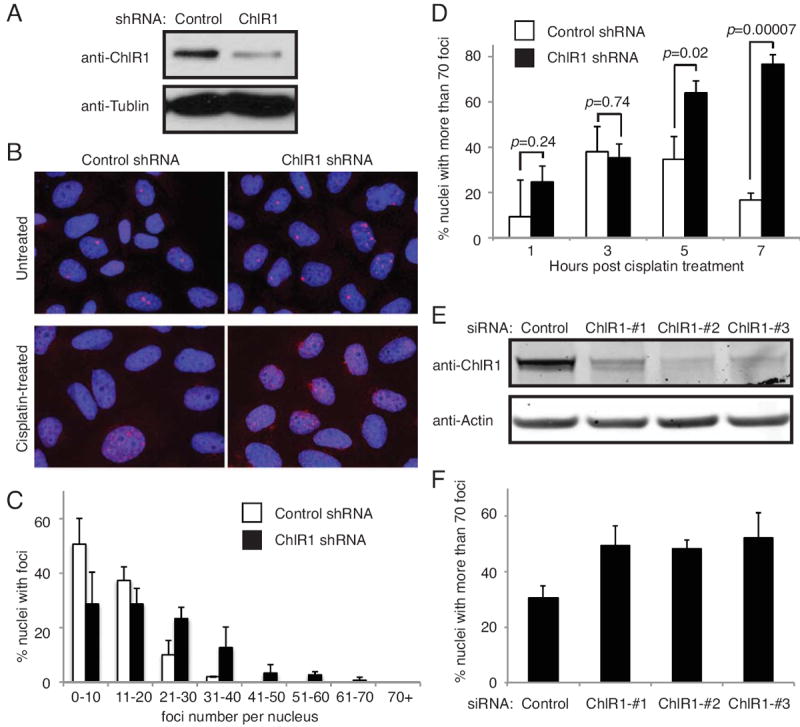
Figure 2. ChlR1 depletion leads to accumulation of 53BP1 foci.

(A) Western blot analysis of HeLa cells stably expressing the indicated shRNAs. (B) Representative images of 53BP1 foci in control and ChlR1-depleted cell nuclei. In the top panel, cells infected with the indicated shRNAs were grown on coverslips and stained with the 53BP1 antibody (red). In the bottom panel, cells were treated with cisplatin for 1 hour and returned to fresh medium for 7 hours. Cells were then stained with the 53BP1 antibody (red). DNA was costained with DAPI (blue). The merged images of DNA and 53BP1 are shown. (C) Quantification of 53BP foci in untreated cells. 53BP1 foci were counted in at least 50 nuclei in each experiment. The number of 53BP1 foci in each nucleus was counted and scored within a category of foci number per nucleus. For each category, an average percentage of nuclei and standard deviation were determined. (D) Quantification of 53BP1 foci in cisplatin treated cells was performed as in C. The number of 53BP1 foci in each nucleus was counted at the indicated time after 1 hour of cisplatin treatment. Percent nuclei with more than 70 foci is shown. (E) HeLa cells were transiently transfected with the indicated siRNA oligonucleotides. Cells were collected 4 days after transfection, and levels of ChlR1 were monitored by Western blotting, using antibody against ChlR1. (F) siRNA-treated cells used in E were incubated with cisplatin for 1 hour and returned to fresh medium without cisplatin for 7 hours. Cells were then stained with the 53BP1 antibody, and cells with more than 70 53BP foci were scored in at least 100 nuclei. Data were obtained from four independent experiments and error bars represent standard deviation.
