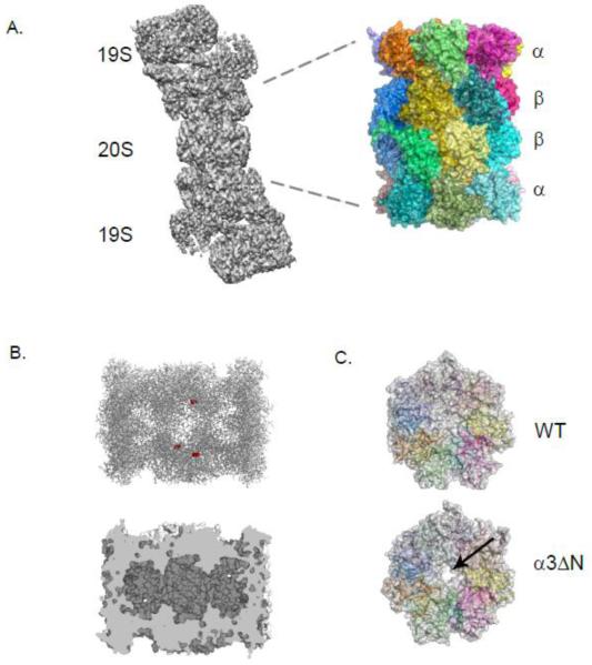
Figure 1. Structural features of the Eukaryotic 20S Proteasome.
A. An electron micrograph structure of the 26S proteasome (grey) from yeast with the 20S (CP) and 19S (RP) labeled. The yeast CP crystal structure is shown with each subunit colored.
B. In the top panel, a clipped structure of the CP on its side illustrates three of the six catalytically active sites shown as red spheres. The bottom panel highlights the three linked chambers within the proteasome.
C. The occluded pore of the CP (grey with individual subunits colored) from wild type cells is juxtaposed with the open pore from the α3ΔN strain indicated by an arrow.
All images were made using PyMol or Chimera using structures elucidated in Refs [15, 29].

