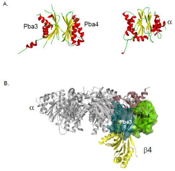
Figure 5. Proteasome biogenesis associated (Pba) factors 3 and 4.
A. The structure of the Pba3/4 dimer is similar to the structure of proteasome subunits. A representative α subunit is shown for comparison. Helices are indicated in red, β strands in yellow and loops in green.
B. The Pba3 (blue) and Pba4 (green) chaperone associates with the α5 subunit (purple) in such a manner that there is a steric clash with the β4 subunit (yellow) unless Pba3/4 disassociates before this subunit is incorporated into the CP.
These images were made using PyMol using structures elucidated in Refs [15, 69].

