
FIGURE 2.
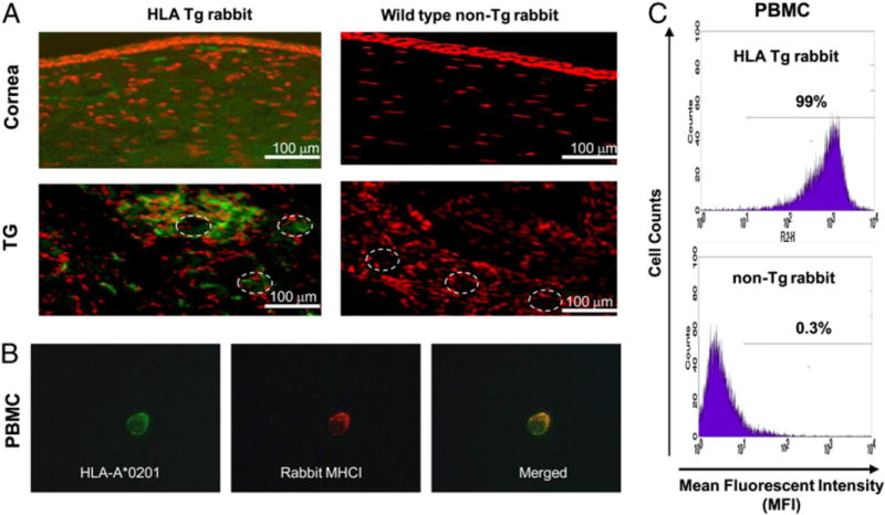
Detection of HLA-A*0201 molecules in cornea, TG, and PBMC of HLA transgenic rabbits. A, Cornea and TG sections from either HLA Tg rabbit or from wild type nontransgenic rabbits (control) were immunostained with FITC (green) conjugated BB7.2 mAb (anti-human HLA-A*0201) and analyzed by fluorescence microscopy (see Materials and Methods). Cell nuclei are shown in red. Dashed circles delineate neuron bodies in the TG (original magnification 320). B, Coexpression of HLA-A*0201 (green) and rabbit MHC class I (red) on the surface of a PBMC derived from an HLATg rabbit and detected by fluorescence microscopy. PBMCs from HLA Tg rabbit were double stained with two different primary Abs HLA-A2.1 mAb (BB7.2) and a rabbit MHCI followed by staining with secondary Abs conjugated with two different fluorescence probes, either Alexa Fluor 594 (green) or Alexa Fluor 488 (red), respectively. Merged yellow signals indicate the colocalization of these two molecules on the surface of PBMCs. C, PBMCs from either HLA Tg rabbit or from wild type nontransgenic rabbits were first stained with BB7.2 mAb, then with PE conjugated anti-mouse secondary IgG Ab, and analyzed by flow cytometry (original magnification ×20).