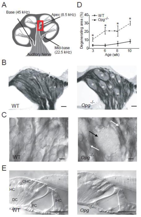
Figure 1. Degenerative changes in the cochlear nerve in OPG deficient mice.
(A) A schematic of a cochlear cross section depicting 3 regions that were studied, and sound frequencies that these regions are tuned to. The boxed region in the apex indicates the spiral ganglion that contains somata of cochlear neurons shown in (B, C) and Fig. 2A. (B, C) Osmicated, plastic-embedded sections of 10 wk old cochlear neurons showed that cell bodies of most WT neurons were individually surrounded by osmiophilic Schwann cells (left) whereas cell bodies of Opg−/− neurons formed degenerating aggregates with poorly defined cellular boundaries (marked with white asterisks in B and black arrow in C). Scale bar: 50 μm (B), 10 μm (C). The white arrows point to normal neurons (C, right). (D) The area of aggregates of degenerating neuronal cell bodies, when expressed as a fraction of the total area occupied by neuronal cell bodies of a cochlear apical half turn was larger and increased faster with age in Opg−/− than in WT mice. The error bars indicate the standard errors of the results from 4–13 ears from 4–10 animals for each age. (E) Osmicated, plastic-embedded sections of 10 wk old WT (left) and Opg−/− (right) mice showed that there was no obvious abnormality in the organ of Corti, which includes sensory inner hair cells (IHC) and outer hair cells (OHC), as well as supporting cells comprising Deiters cell (DC), pillar cell (PC) and Hensen’s cell (HC). Scale bar: 25 μm.

