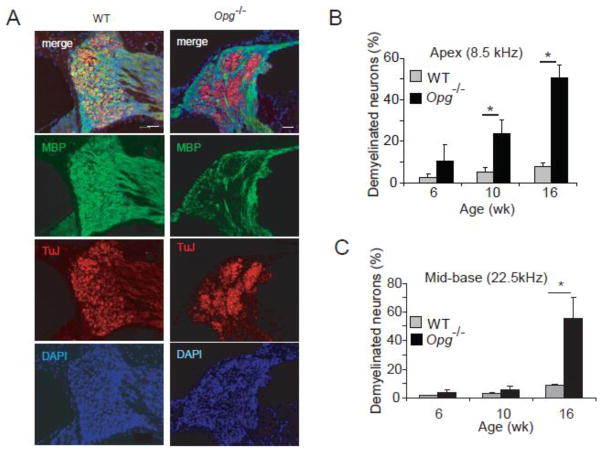
Figure 2. Loss of myelination in OPG deficient cochlea.
(A) Immunohistochemistry for TuJ and MBP at 10 wk of age confirmed demyelination of pathologic neural aggregates. DAPI is a nuclear stain. Scale bar: 50 μm. (B) The fraction of demyelinated neurons compared to the total number of neurons significantly increased with age in Opg−/− compared to the age-matched WT mice in the cochlear apex. This fraction refers to the Tuj positive neurons that are not surrounded by the MBP signal compared to TuJ positive neurons that are surrounded by the MBP signal. (C) The fraction of demyelinated neurons compared to the total number of neurons significantly increased with age in Opg−/− compared to age-matched WT mice in the cochlear base.*P<0.05. For bar graphs, gray indicates WT and black Opg−/− in this and other figures. Data expressed as mean +/− standard error of the mean in all figures. The error bars in (B) and (C) are based on measurements from 3 different animals per group.

