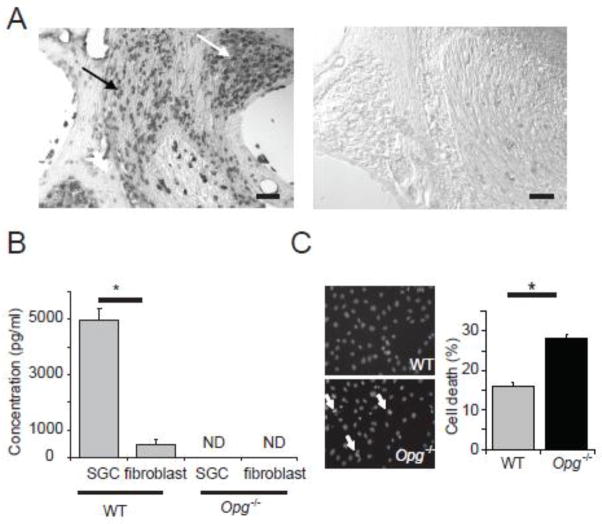
Figure 4. Expression and secretion of OPG by cochlear neurons and Schwann cells.
(A) In situ hybridization for Opg showed strong signal in cochlear neurons (white arrow) and Schwann cells (black arrow) in WT cochlea (left), and no signal with the control (sense) probe (right). Scale bar: 100 μm. (B) Measurements of secreted OPG in culture medium of WT SGC and fibroblasts showed that OPG was abundantly secreted by WT SGC but not secreted by Opg−/− cells. The error bars indicate the standard errors of three replicates. ND: not detectable. (C) Treatment of cultured SGC with H2O2 resulted in more nuclear condensations in Opg−/− cells than in WT cells, as shown in the representative images (left, white arrows), and bar graphs (right); cells in 10 random fields in each independent experiment were counted. The error bars indicate the standard errors of 4 replicates. The nuclei were stained with Hoechst 33342.

