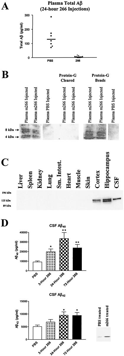
Figure 2.
m266 sequesters Aβ in vivo. (A) Three-month-old PDAPP+/+ mice were treated with PBS (n = 7) or 500 μg of m266 (n = 9) i.v. Twenty-four hours after m266 administration, plasma was analyzed for AβTotal by RIA. Before analysis, all plasma samples were first treated with protein G to remove Aβ complexed to m266. (B) In the first three lanes, 20 μl of plasma from PDAPP mice (24 h after administering 500 μg of m266 or PBS i.v.) was run on acid-urea gels followed by Western blotting for Aβ with m6E10 (Senetek, Napa, CA). Lanes 4 and 5 demonstrate that the plasma Aβ, from 20 μl of m266-injected mice, can be completely cleared with protein G treatment for animals treated with m266. In lanes 6 and 7, the protein-G beads from the 20 μl of plasma from lanes 4 and 5 were washed, denatured in formic acid, and analyzed. Lane 8 represents the protein G beads from 20 μl of plasma from a PBS-injected mouse. (C) Tissues collected from adult PDAPP+/+ mice were homogenized in a buffer containing 1% SDS. One hundred micrograms of total protein from each lysate was subjected to reducing 8% SDS/PAGE and analyzed by Western blotting for human APP with m6E10. (D) CSF Aβ40 and Aβ42 was determined by RIA from 3-month-old PBS- (n = 23) and m266-treated PDAPP mice 3 (n = 9), 24 (n = 5), and 72 h (n = 9) after i.v. injection of m266 as above. There was significantly greater Aβ40 in the CSF of the m266-treated mice at 3, 24, and 72 h and Aβ42 at 24 and 72 h (*, P < 0.05; **, P < 0.0001, ANOVA followed by post hoc Tukey's test.). Two microliters of CSF from each mouse per treatment group was collected, pooled (15 μl total), and subjected to acid urea gels followed by Western blotting with Aβ-specific antibodies.

