
Figure 3.
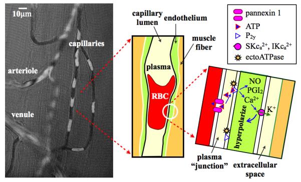
Figure illustrates the potential for direct transfer of ATP from the erythrocyte (RBC) to P2y receptors on the capillary endothelium. Left panel shows composite image of a single video frame (opacity 64%) superimposed on a functional (minimum) image from the same video. Video sequence captured at 40X with a 1-msec exposure using a Rolera XR digital video camera on an Olympus IX-81 inverted microscope using a 420-nm interference filter. The video frame has been processed as a pseudo-optical density image with low to high values scaled from black to white. The functional image (29) represents the minimum light intensity value at each pixel over the video sequence and hence shows the capillary lumen as defined by the passage of erythrocytes (glycocalyx not included). In the composite image, white erythrocytes are superimposed on the black capillary lumen. The middle panel shows a schematic of a single erythrocyte in the capillary and the right panel a schematic of a magnified view of the plasma junction between erythrocyte and endothelial membranes. ATP released from the erythrocyte via pannexin 1 channels diffuses across the narrow gap to P2y receptors on the capillary endothelium inducing the synthesis and release of nitric oxide (NO) and prostacyclin (PGI2) as well as Ca2+ signaling in the endothelium resulting in hyperpolarization of the endothelial cell via Ca2+ sensitive K+ channels.