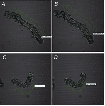
Figure 11. The absence of AC6 impairs the secretory response to VIP in isolated duct fragments.

A and B, a representative duct fragment before (A) and after VIP stimulation (10 nm) (B) from WT mice. C and D, a representative duct fragment before (C) and after VIP stimulation (D) from AC6−/− mice. The lumen area is drawn in green. An expansion in lumen area is observed in sealed duct fragments from WT mice (area: 16267.03 μm2 vs 20993.34 μm), but not from AC6−/− mice (area: 5124.47 μm2 vs 4996.56 μm2).
