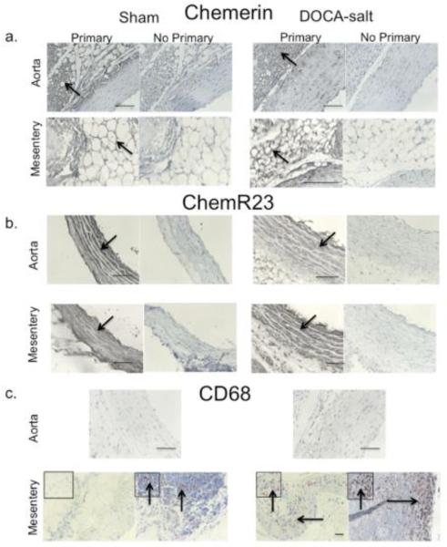
Figure 4. The Chemerin Axis in DOCA-salt Hypertension.
Immunohistochemical staining of chemerin (a), ChemR23 (b) and CD68 (c) in thoracic aorta and superior mesenteric artery from the sham normotensive (left) and DOCA-salt hypertensive rat (right). Representative of 4-5 different animals for each group. Arrows are regions of interest with boxed insets magnified. Bars = 100 microns.

