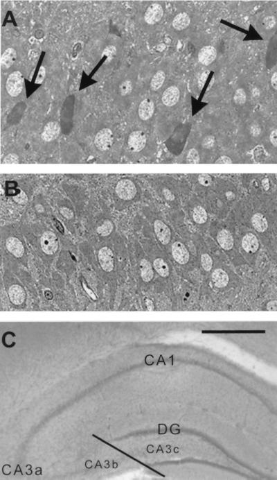
Figure 2.
Acute injury of CA3 hippocampal pyramidal cells in P13 rats results from CRH administration. (A) Shrunken, toluidine blue-stained, injured cells (arrows) are visible in 1-μm sections from CRH-treated rats, but not in sections from vehicle-treated controls (B). (C) Subdivisions of CA3 pyramidal cell layer, denoting the CA3b/CA3c border. [Scale bars = 20 μm (A and B) and 200 μm (C).]

