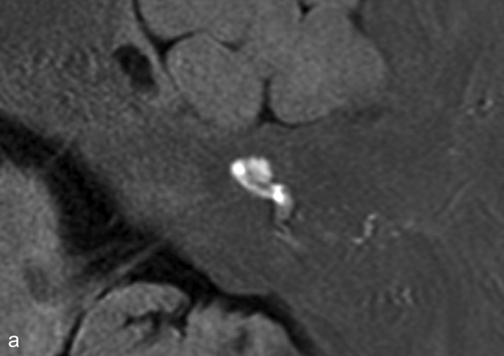
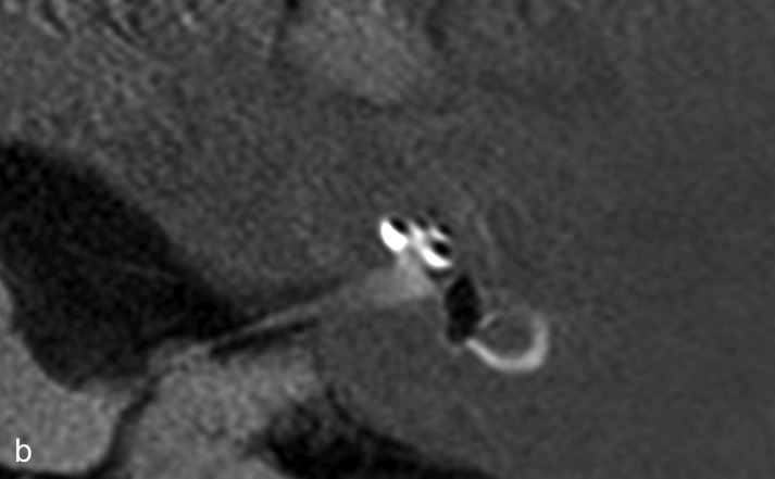
Figure 1.
Endolymphatic hydrops as seen on high-resolution magnetic resonance imaging of the petrosal bone 24 h after transtympanic injection of gadolinium, which diffuses predominantly into the perilymphatic space.
a) The labyrinth of a healthy control: The cochlea and semicircular canals are visualized.
b) The labyrinth of a patient with Menière’s disease: the endolymphatic hydrops can be recognized by virtue of its lack of contrast medium uptake.
With kind permission by Robert Gürkov


