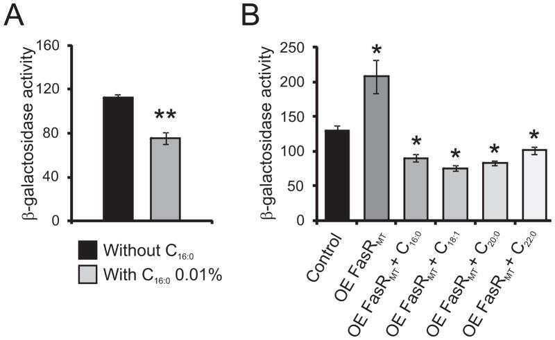
Figure 7.
A. Intracellular β-galactosidase activity of strain MSpFR47 grown in 7H9 medium with or without C16:0 0.01 %. Levels of activity are shown as nmol ONPG per min per mg of protein and are the means of the results of three independent experiments ± standard deviations (n=3). **, P = 0.015.
B. Intracellular β-galactosidase activity of strain MSpFR47 pFR9 grown in 7H9 medium without acetamide (control), MSpFR47 pFR9 grown in 7H9 medium with acetamide 0.2% (OE FasRMT) and MSpFR47 pFR9 grown in 7H9 medium with acetamide 0.2% and in the presence of different fatty acids at a final concentration of 0.01 % (OE FasRMT + C16:0 to OE FasRMT + C24:0). Samples of each culture were removed 4 h post-induction to assay β-galactosidase specific activity. Levels of activity are shown as nmol ONPG per min per mg of protein and are the means of the results of three independent experiments ± standard deviations (n=3). *, P <0.0001.

