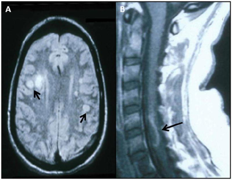
Fig. 1. MRI scans of patients with varicella zoster virus (VZV) multifocal vasculopathy and myelopathy.

(A) Proton-density brain MRI scan shows multiple areas of infarction in both hemispheres, particularly involving white matter. Arrows point to gray-white matter junction lesions. (Reproduced from Gilden et al. [107]; copyright 2002, Springer; with permission.) (B) Note cervical, longitudinal, serpiginous enhancing lesions (arrows). (Reproduced from Gilden et al. [81]; copyright 1994, Wolters Kluwer Health; with permission.)
