
Fig. 3. Varicella zoster virus (VZV) in the temporal artery of a patient with VZV multifocal vasculopathy.
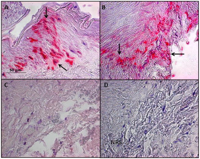
(A) Positive control cadaveric cerebral artery 14 days after VZV infection in vitro (pink color, arrows). (B) Note VZV antigen in the adventitia of the temporal artery after staining with anti-VZV antibody (pink color, arrows), but not after staining adjacent sections with anti-HSV-1 antibody (C) or normal rabbit serum (D). Magnification 200X. (Reproduced from Mathias et al. [110]; copyright 2013, Elsevier; with permission.)