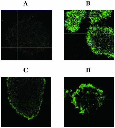
FIG. 6.
Scanning confocal photomicrographs of P. aeruginosa biofilms established in flow cells. Select microcolonies stained with ConA-FITC, which reveals polysaccharides such as alginate, are shown from the top. (A) PAO1 biofilm not exposed to antibiotics; (B) PDO300 (a derivate of PAO1 that constitutively overproduces alginate) not exposed to antibiotics; (C) PAO1 exposed to imipenem for 18 h; (D) PAO1 exposed to imipenem for 37 h.

