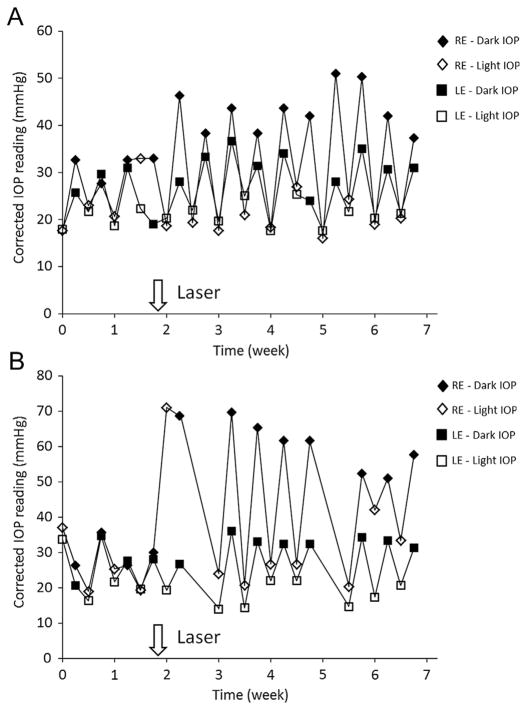
Fig. 2.
Representative IOP profile after partial trabecular laser photocoagulation. (A) Laser photocoagulation was applied at 270° trabecular meshwork. The dark phase IOP was significantly elevated after laser treatment for 5 weeks but there was no significant increase in light phase IOP compared to contralateral control eye. The IOP profile of animal #1712 is shown. (B) Laser photocoagulation was applied at 330° trabecular meshwork. The light phase IOP was dramatically increased in the first week after laser photocoagulation and returned to the IOP level comparable to the contralateral untreated control eye. The dark phase IOP was markedly increased. The IOP profile of animal #1730 is shown. Consistent circadian pattern of light and dark phase IOP was noted in untreated control eye. RE = right eye with laser treatment; LE = left eye as contralateral control.

