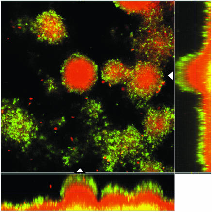
FIG. 5.
Epifluorescence and scanning confocal laser photomicrographs of PAO1-J32 biofilms on day 6 showing the activity of the ampC promoter in response to ceftazidime exposure (100 μg/ml for 4 h). The micrograph consists of a horizontal section and two vertical sections through the biofilms collected at the positions indicated by the white triangles in the horizontal section. The position of the horizontal image is indicated by the crossing lines in the vertical sections. The biofilms were stained red with SYTO 62, demonstrating the presence of biomass in the flow cell. The expression of Gfp indicates the expression of the AmpC β-lactamase in the biofilm.

