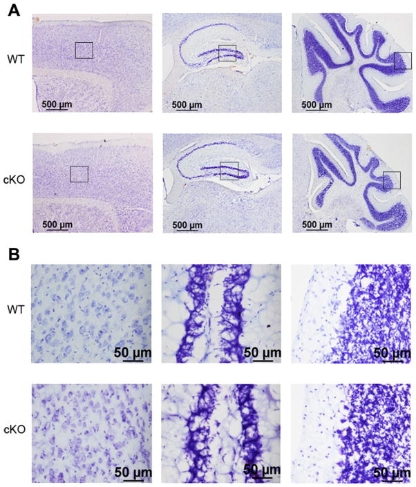
Fig. 4.
Histological analysis of Nes-Cre/HDAC4 cKO mice. Sagittal sections stained with cresyl violet are shown from a 9-week-old Nes-Cre/HDAC4−/− (cKO) and wild-type (WT) littermate. A: Pictures from three brain regions are displayed at low magnification, from left to right: cortex, hippocampus, and cerebellum. B: Higher magnifications of the same brain regions from A. [Color figure can be viewed in the online issue, which is available at wileyonlinelibrary.com.]

