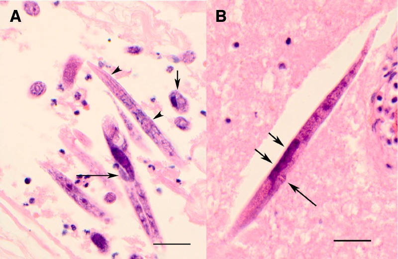
Figure 1.
Photomicrographs of brain tissue containing Halicephalobus sp. A, Numerous sections of females and larvae, showing rhabditoid esophagus (arrowheads), flexed ovary (long arrow), and a cross section (short arrow) showing intestine (on the right) and single reproductive tube (on left), (H&E stain, ×600). B, Longitudinal section of a female worm showing protuberant vulva (long arrow) and two eggs (short arrows) (Hematoxylin and eosin stained, original magnification ×600, scale bars = 50 μm).

