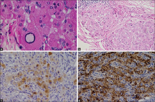
Figure 4.

Permanent histopathological and immunohistochemical evaluation: (a: upper left) Cells contain lipochrome pigments and show mild nuclear pleomorphism with absence of Reinke crystals and lack of mitotic activity (H and E, ×400), (b: upper right) Fibrosis surrounding the eosinophilic polygonal cells and lymphoid aggregates (H and E, ×200), (c: lower left) Patchy reactivity for synaptophysin (IHC, ×200), (d: lower right) Positive reactivity to CD56 (IHC, ×200)
