Figure 1.
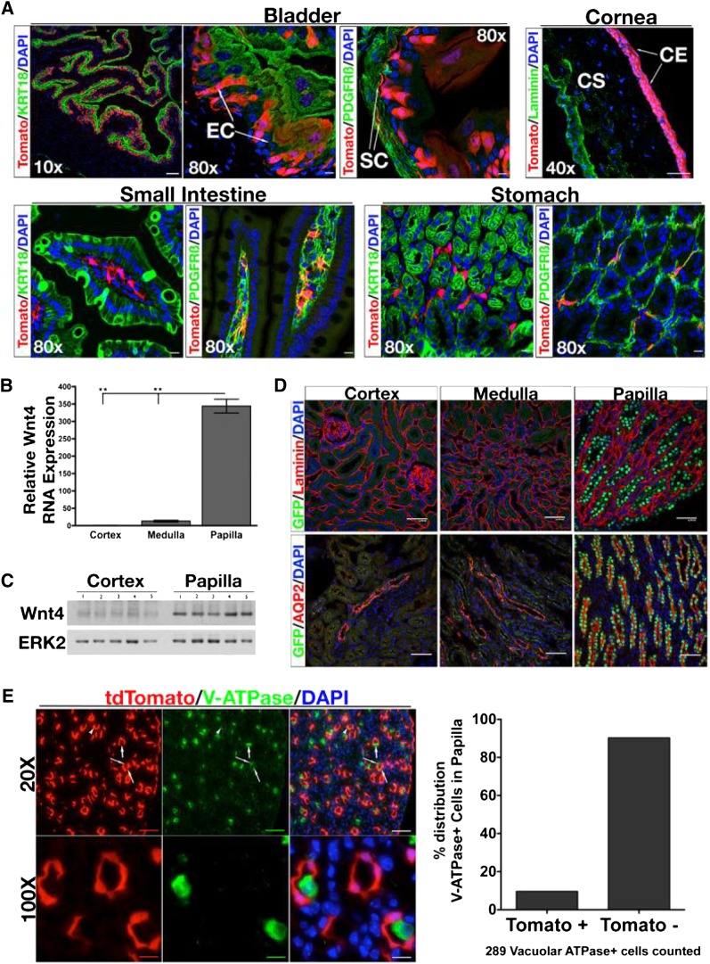
Validation of Wnt4GCE/+;R26tdTomato/+ reporter mice. (A) tdTomato reflects Wnt4 expression. tdTomato+ cells are seen in epithelial KRT18+ cells, and stromal PDGFRβ+ cells of the bladder. tdTomato is strongly detected in corneal epithelium. We also report tdTomato+ cells in tissues that have not been previously reported to express Wnt4. In the small intestine and stomach, tdTomato+ cells colocalize with PDGFRβ+ cells. (B) Cortex, medulla, and papilla are dissected from wild-type C57BL/6N mice, RNA is extracted, and SybrGreen-based qPCR is performed to detect Wnt4. Wnt4 transcript is significantly upregulated in papilla versus cortex and medulla. (C) Whole cell protein homogenate from cortical and papillary samples are processed for Western blot to test for Wnt4 protein levels. (D) Confocal images of renal cortex, medulla, and papilla from Wnt4GC/+ reporter mice. In the top row, sections are stained with anti-laminin antibody (red), anti-GFP antibody (green), and DNA marker DAPI (blue). Nuclear GFP represents Wnt4+ cells, which are not detected in cortex and medulla but are detected in papilla tubules. The images in the bottom row are of kidney sections stained with anti-aquaporin 2 antibody (red), anti-GFP antibody (green), and DAPI (blue). GFP+ cells colocalize with aquaporin 2+ cells in the papilla and not in the cortex or medulla, indicating that Wnt4+ cells are located specifically in papillary CD epithelia. (E) Sections of papilla from Wnt4GCE/+;R26tdTomato/+ stained with anti-V-ATPase to identify intercalated cells. Quantification reveals that >90% of V-ATPase+ cells are tdTomato−. In B, n=3 or 4 per group. Data analyzed by one-way ANOVA. **P<0.0001 (Tukey’s multiple comparison test). EC, epithelial cell; SC, stromal cell; CE, corneal epithelium; CS, corneal stroma; DAPI, 4',6-diamidino-2-phenylindole. Scale bars, 100 µM in ×10 images in A; 50 µM in ×40 images in A; 10 µM in ×80 in A.

