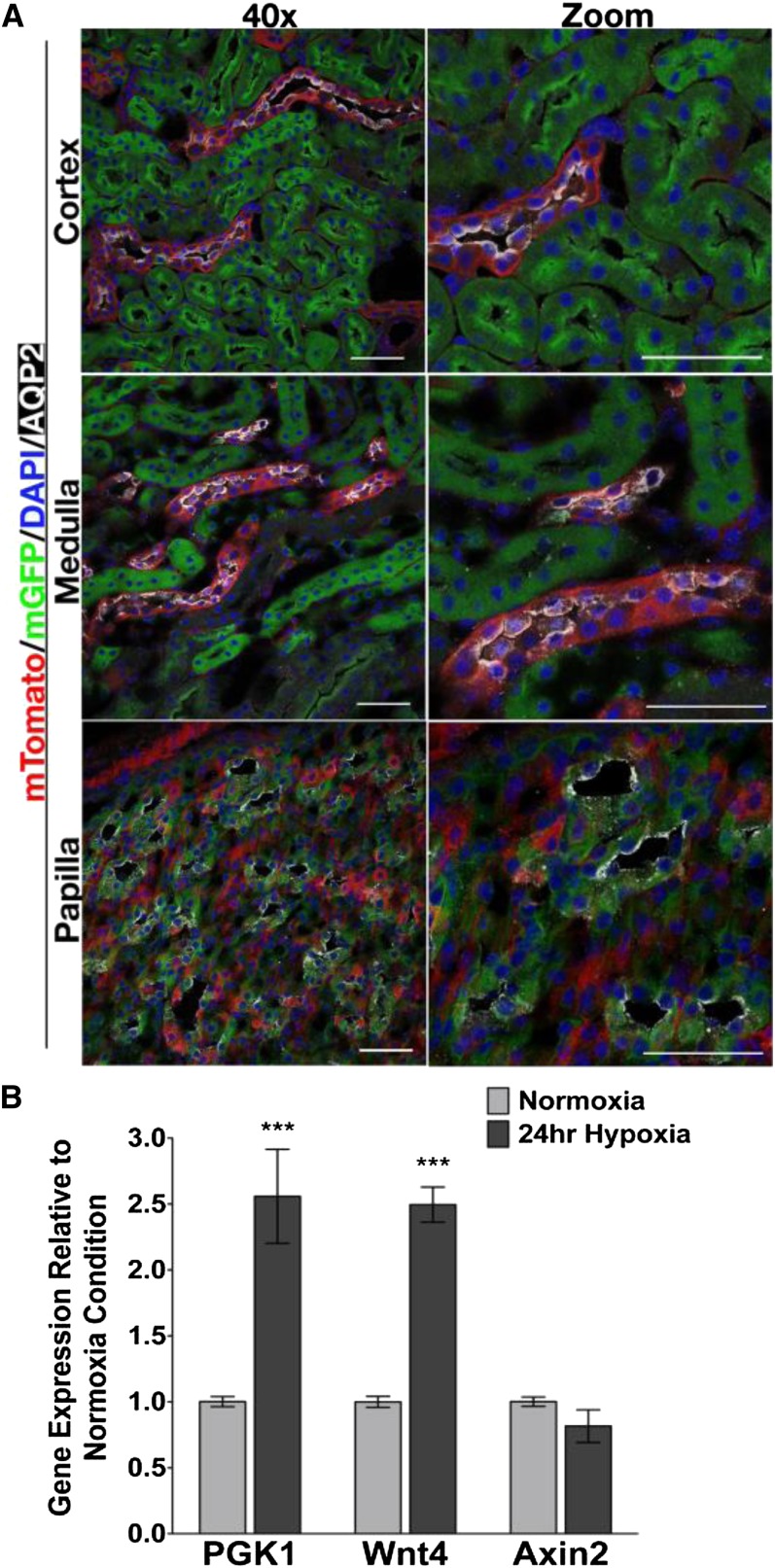
Figure 2.
Wnt4 is not expressed in cortical collecting duct during development. (A) Confocal images of cortex, medulla, and papilla taken with a ×40 objective lens in the left column and high-power digital zoom in the right column. Kidney sections are from Wnt4GC/+;R26mTmG/+ mice. Green cells indicate that Wnt4 is expressed in that cell or has been previously expressed in a progenitor of that cell. Red cells never expressed Wnt4. Red and green fluorescence is epifluorescence, blue is DAPI, and white is AQP2. In cortex and medulla, aquaporin 2+ CD epithelia are in red cells. In papilla, AQP2+ cells are in green (Wnt4 expressing or descendant) cells. (B) Primary IMCD cells are grown in normoxic (20% O2) or transiently exposed to hypoxic (3% O2) for 24 hours. Gene expression as measured by qPCR indicates that the hypoxia-responsive gene PGK1 is significantly increased in hypoxia exposed IMCD cells. Wnt4 gene expression is also significantly increased in hypoxic IMCD cells compared with normoxic cells, whereas Axin2 gene expression is unchanged. Data are analyzed by two-way ANOVA comparing gene expression between hypoxic and normoxic conditions (Bonferroni post test). ***P<0.001. Scale bars, 50 µM in A. DAPI, 4',6-diamidino-2-phenylindole; AQP2, aquaporin-2; IMCD, inner medullary collecting duct.

