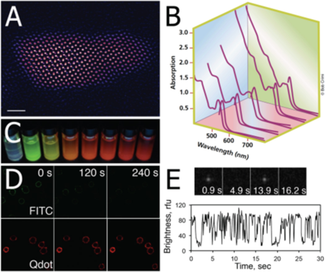
Figure 1.
Photophysical properties of quantum dots. (A) Aberration-corrected Z-STEM image of a commercial core/shell QD655 (Invitrogen). It can be seen that the core/shell QD is actually an elongated, bullet-shaped 3D object rather than a sphere. (Reprinted with permission from Ref 7. Copyright 2007 Elsevier B.V.) (B) Absorption and emission spectra of a series of CdSe nanocrystals. The size of the QD core determines the absorption and emission spectra of QDs. (Reprinted with permission from Ref 14. Copyright 2001 Nature Publishing Group) (C) A series of UV-illuminated CdSe nanocrystals ranging in size from ~ 2 nm to ~6 nm. (D) Time-lapse image series of FITC- and QD-labeled HEK-293 cells. In contrast to traditional fluorophores, QDs are characterized by excellent photostability which enables long-term monitoring of biological processes. (Reprinted with permission from Ref 48. Copyright 2011 American Chemical Society) (E) Fluorescence intermittency or “blinking” in the emission spectrum of a single QD. This QD property can be used as a criterion to distinguish single nanocrystals from aggregates.

