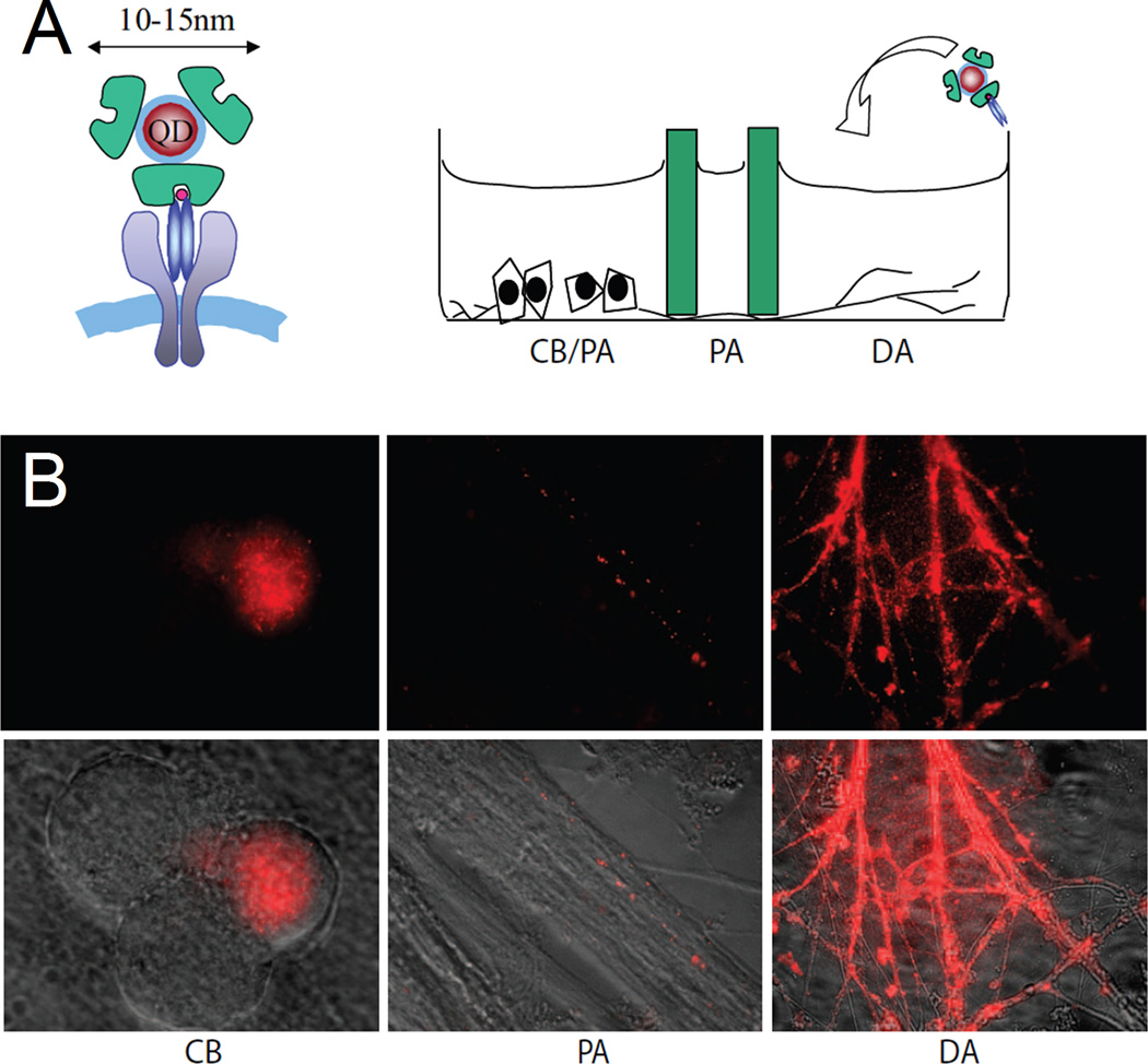
Figure 6.
QDot labeling of NGF receptors using Streptavidin-biotin assembly. NGF-conjugated QDs were used to investigate retrograde, vesicular NGF transport in living DRG neurons. (A) Schematic drawing of a QDot-NGF bound to dimerized TrkA receptors (Left) and addition of QDot-NGF to the DA compartment of the three-chamber DRG neuron culture (Right). DA, distal axon; PA, proximal axon; CB, cell body. (B) Representative live fluorescence images of DRG neuron axons or cell bodies 2 h after the addition of 4 nM QDot-NGF to the DA chamber. QDot-NGF seems to bind all axons in distal axon chamber. However, only a small portion of the cell bodies and proximal axons are shown to have QDot fluorescence, reflecting the fact that not all cell bodies extend their axons into the distal axon compartment. (Reprinted with permission from Ref 63. Copyright 2007 National Academy of Sciences, U.S.A.)

