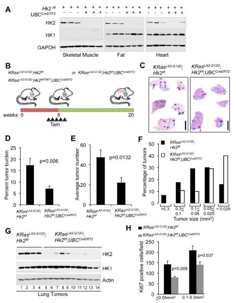
Figure 6. Systemic deletion of Hk2 in the mouse is therapeutic for oncogenic KRas-driven NSCLC.
A. Immunoblot showing HK2 and HK1 protein expression in tissues isolated from either Hk2f/f or Hk2f/f;UBCCreERT2 mice two weeks after final tamoxifen injection. B. Schematic illustration of the experiment described in C–H. C. Representative images of H&E staining of whole-mount lung sections prepared from either KRasLA2-G12D;Hk2f/f or KRasLA2-G12D;Hk2f/f;UBCCreERT2 lungs, 12 weeks after injection of tamoxifen at 8 weeks of age. Individual tumors are indicated by arrows (scale bar 4mm). D. Percentage tumor burden as calculated from the H&E sections of tumor bearing lungs from either KRasLA2-G12D;Hk2f/f or KRasLA2-G12D;Hk2f/f;UBCCreERT2 mice at 20 weeks of age and after injection of tamoxifen at 8 weeks of age (n=8 per group). E. Tumor numbers calculated from the H&E stained sections of tumor bearing lungs from either KRasLA2-G12D;Hk2f/f or KRasLA2-G12D;Hk2f/f;UBCCreERT2 mice (n=8 per group). F. Size distribution of tumors developed in the lungs of either KRasLA2-G12D;Hk2f/f or KRasLA2-G12D;Hk2f/f;UBCCreERT2 mice (n=6 per group). G. Immunoblot showing HK2 and HK1 protein levels in tumor lysates extracted at 20 weeks of age, and after tamoxifen injection at 8 weeks of age. Lanes 1–4- tumors were randomly selected for analysis; lanes 5,6 – tumor size < 0.025mm2; lanes 7–9- tumor size 0.3–0.05mm2; lanes 10–14-tumor size 0.05–0.025mm2. H. Quantification of Ki67 staining of the same size groups of tumors derived from either KRasLA2-G12D;Hk2f/f or KRasLA2-G12D;Hk2f/f;UBCCreERT2 mice. Data presented in Figs. D, E and H are expressed as the mean ± SEM. See also Fig. S4.

