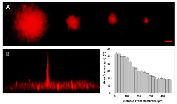
Fig. 5.
Confocal images of MI-Texas Red in bulk CNBC-A following UV irradiation with circular photomasks. A) Top view of summed voxel projection of MI-Texas Red bound through depth of CNBC-A using 10-pixel, 8-pixel, 6-pixel, and 4-pixel diameter circle photomasks, from left to right (scale bar = 50 μm). B) Side view of 3D image stack of MI-Texas Red binding after using the 4-pixel mask, with binding depth of 472 μm. C) Graph of the mean diameter of MI-Texas Red binding through depth of CNBC-A from Part B, where the cell culture insert membrane is at 0 μm and bar width is equivalent to summed slice thickness (error bars represent standard deviation)

