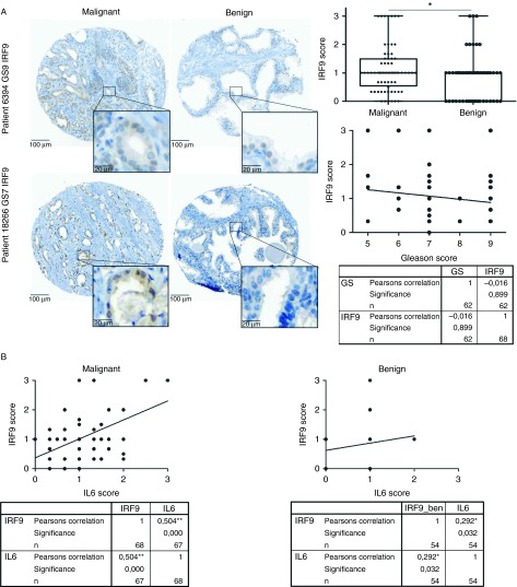
Figure 3.
IRF9 expression in human prostate tissue. (A) Immunohistochemical staining of one representative benign and one malignant core from two prostate cancer (PCa) patients for IRF9 and evaluation of staining intensity of the PCa TMA. (B) Immunohistochemical staining of IL6 was performed on a consecutive slice of the TMA. Expression of IL6 was correlated with that of IRF9 in benign and malignant cores of PCa patients. *P<0.05; **P<0.01.

