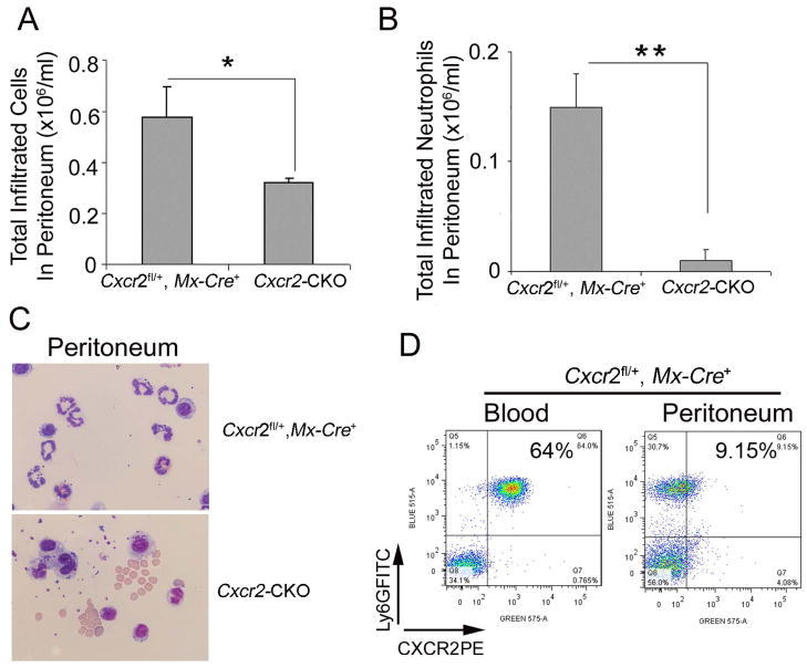
Figure 6. Decreased migration of CXCR2-deficient neutrophils in sterile peritonitis.
Cxcr2-CKO and Cxcr2fl/+::Mx-Cre+ mice 4 wks after poly(I:C) injection were injected with 4% aged TG 2 hours before analysis. (A) Total cells were collected from the peritoneum and counted on a hemocytometer. (B) Total infiltrated neutrophils in peritoneum were calculated by total cells collected from the peritoneum times the percentage of neutrophils in total cells determined by the staining of peritoneal cells with Ly6G and CD45 antibodies (data not shown). (C) Wright-Giemsa staining of peritoneal cells collected by cytospin showed neutrophils with multiple lobulated nuclei in the peritoneal exudate of Cxcr2fl/+::Mx-Cre+mice (top) and mononuclear and kidney-shaped nuclei consistent with monocytes in the peritoneum of Cxcr2-CKO mice (bottom). (D) Peritoneal exudate cells and blood cells from Cxcr2fl/+::Mx-Cre+ mice were collected and stained with Ly6G and CXCR2 antibodies, then analyzed by flow cytometry. These data represent two independent experiments. Each experiment included 3 mice per group. **P<0.01; *P<0.05;

