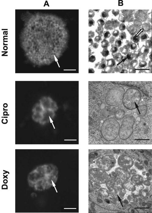
FIG. 2.
Immunofluorescence and electron microscopy of inclusion morphology in the presence and absence of persistence-inducing antibiotics. C. pneumoniae inclusions grown in the presence or absence of antibiotics for 72 h postinfection were visualized by confocal immunofluorescence (column A) or electron (column B) microscopy. Confocal microscopy of normally grown C. pneumoniae revealed large inclusions with granular staining structures (white arrow) that corresponded to EBs (black arrow) visible in a similar sample visualized by electron microscopy. Only few regular-size RBs are visible (white arrow). In contrast, doxycycline (Doxy)- and ciprofloxacin (Cipro)-treated cultures exhibited altered morphology at 72 h postinfection. Inclusions were smaller. Enlarged RB structures were evident in both confocal (white arrows) and electron (black arrows) microscopy micrographs of these samples. EBs were lacking in the antibiotic-treated samples, and the visible RBs were roughly three to five times the size of regular RBs under normal growth conditions. Scale bars: A, 2 μm; B, 1 μm.

