
Fig. 1.
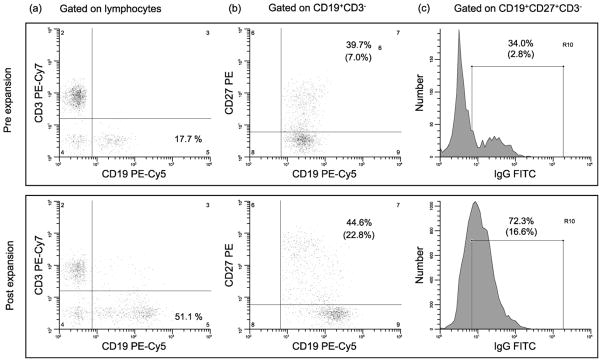
Representative flow cytometric analysis pre- and post-expansion. Staining of PBMC performed before and after expansion in a representative volunteer; gated on the lymphocyte region as defined by forward versus side light scatter (a); gated on CD19+ CD3− (b); gated on CD19+ CD27+ CD3− (c); in parentheses under the percentages of cells in selected regions are the % of positive cells in the corresponding region as related to the number of cells in the lymphocyte region.