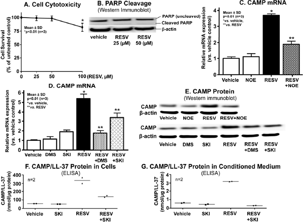
Figure 1.
RESV-mediated increase in S1P is responsible for stimulation of CAMP expression. HaCaT KC pretreated with or without ceramidase (NOE, 25 µM) or SPHK (DMS, 2.5 µM; SKI, 1 µM) inhibitors for 30 mins were incubated exogenous RESV (50 µM or as indicated) for 24 h. Cell viability (A) or PARP cleavage as a measure of apoptosis (B). CAMP mRNA expression assessed by qRT-PCR (C and D). CAMP and LL-37 (an active form of CAMP) protein/peptide levels quantified by Western immunoblot analysis (E) and ELISA, respectively (F and G). Similar results were obtained when the experiment was repeated (triplicate) using different cell preparations.

