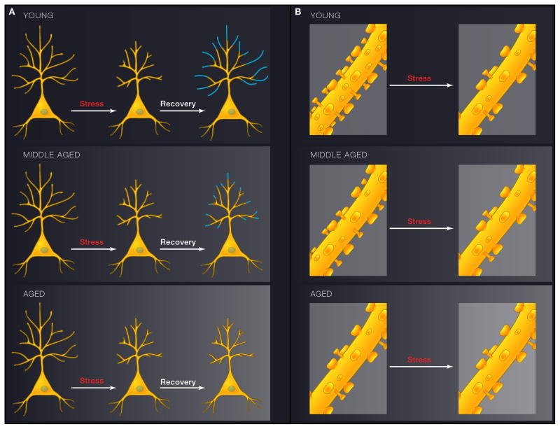
Figure 3.
Schematic diagrams depicting the interactive effects between stress and aging on layer 3 pyramidal neurons in the prelimbic area of mPFC. 3A (left) shows the effects on dendritic arbor and 3B (right), shows the effects on spines. In both cases the upper panel represents young male rats, middle panel represents middle-aged rats, and the bottom panel represents aged rats. A) In young animals, chronic stress leads to shrinkage of distal apical dendrites. After cessation of chronic stress, dendritic trees regrow. Such recovery after stress cessation is blunted by middle age and gone in the aged animals. B) Spines are also lost in young animals exposed to chronic stress, and it is primarily the thin spines that are affected. No further spine loss is induced by stress in middle-aged or aged rats, and this is likely due to the fact that age on its own leads to a loss of the thin spine class (See text for details)

