
Figure 2. Loss of ASIC1a and NaHCO3 administration reduce FPI-induced neuronal degeneration.
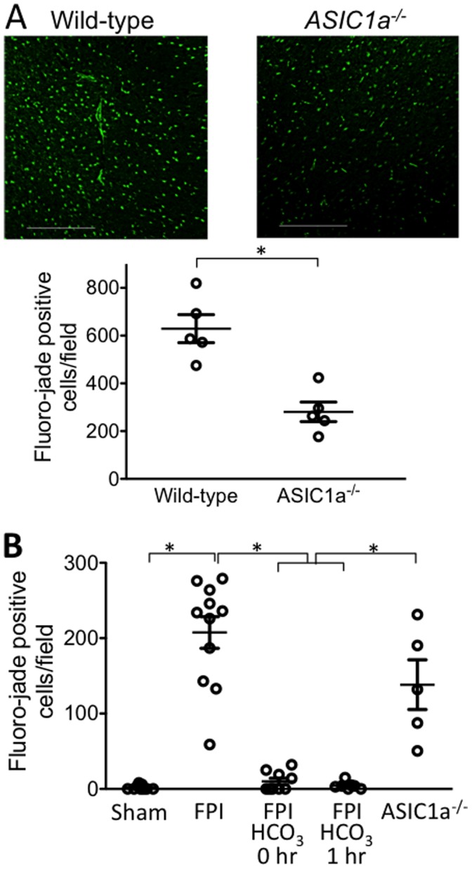
A. Brain was removed one day after FPI. Data are fluoro-jade positive neurons per microscopic field in the cortex (0.9 μm2). N = 5 wild-type and 5 ASIC1a−/− mice. Scale bar = 0.1 mm. Each data point indicates an individual animal. * indicates P<0.05, unpaired t-test. B. Brain was removed four days after FPI. N = 9 sham-treated mice, 11 FPI-treated wild-type mice 3 of which received NaCl after FPI, 9 FPI-treated mice with NaHCO3 administered immediately after FPI, 7 FPI-treated mice with NaHCO3 administered 1 hr after FPI, and 5 FPI-treated ASIC1a−/− mice. * indicates P<0.05, ANOVA with Bonferroni post-hoc test.