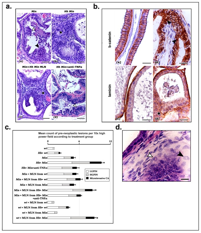
Figure 1. Clinically silent immune system alterations affect dorsolateral prostate (DLP) carcinogenesis in B6 ApcMin /+ mice after GI infection with H. hepaticus.
A) By the age of 12 weeks, Apc Min/+ mice spontaneously develop focal low and high grade prostate intraepithelial neoplasia (LGPIN, white arrow-head; HGPIN, black arrow-head) but only rare microinvasive carcinoma (a). Aged-matched Apc Min/+ mice with H. hepaticus, develop significantly more PIN and microinvasiveCA lesions (asterisk) characterized by highly atypical prostate gland epithelial cells which break through the basal lamina and infiltrate the adjacent stroma (b). When mesenteric lymph node cells from H. hepaticus-infected Apc Min/+ mice are transferred into the peritoneal cavity of H. hepaticus-free Apc Min/+ mice the recipient mice develop preneoplastic and early neoplastic lesions (microinvasiveCA-asterisk) comparable to those found in donor mice (c). Depleting TNF- α from H. hepaticus-infected Apc Min/+ mice brings prostate neoplastic lesions (LGPIN, white arrow-head) occurrence to the level of H. hepaticus-free mice (d). Hematoxylin and Eosin. Bars=50 μm. B) Aberrant β-catenin and laminin immunostaining pattern in microinvasiveCA lesions in prostate cancer. DLP from WT H. hepaticus-positive mice (a and c), H. hepaticus-infected Apc Min/+ mouse (b) and H. hepaticus-free Apc Min/+ mouse transferred with mesenteric lymph node cells from H. hepaticus-infected Apc Min/+ donor (d). Normal DLP glands had a normal β-catenin lateral epithelial cell membrane staining pattern (a) and an intact basal lamina (c). In contrast, microinvasiveCA lesions were characterized by cytoplasmic and nuclear (arrow-heads) stabilization of β-catenin (b) and absence of laminin (asterisk) suggestive of basal membrane degradation in malignant epithelial cell foci of incipient invasion. Hematoxylin counterstain, DAB chromogen. Bars=25 μm. C) Bar graph showing frequency of PIN and microinvasiveCA in treatment groups. The most significantly elevated prostate cancer occurrence is denoted by asterisk. Standard error bars correspond to microinvasiveCA statistical comparisons. D) DLP of H. hepaticus-free Apc Min/+ mouse transferred with MLN cells from a H. hepaticus-infected Apc Min/+ donor, HGPIN. Most myeloid precursor cells with ring-shaped nuclei residing in the stroma have the typical granulocytic lineage phenotype (black arrow-head) while fewer appear to be myelo/monocytic in type (white arrow-head). Bar=16 μm.

