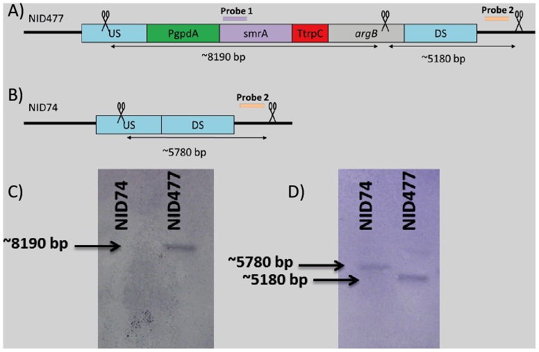
Figure 2. Confirmation of correct integration of smrA in IS1.
A) and B): Schematic overview of the HindIII cut sites (indicated with scissors) and the size of the resulting fragments. Purple and orange bars indicate hybridization site for smrA probe and locus probe respectively. C): Illustration showing placement of the bands relative to each other. D): Southern blot of NID74 and NID477 digested with HindIII and hybridized with smrA probe. E): Southern blot of NID74 and NID477 digested with HindIII and hybridized with locus probe. The illustration is not drawn to scale.

