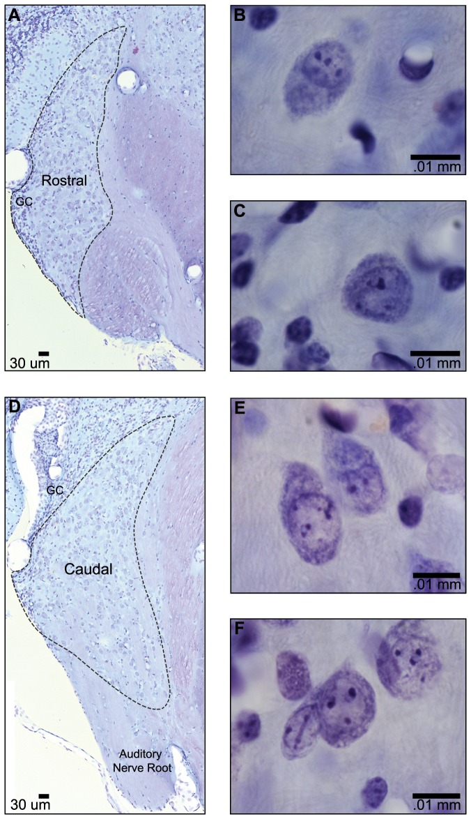
Figure 1. Photomicrographs of Nissl-stained AVCN.
(A) Section through rostral AVCN at low magnification. Granule cells (GC) are present along the lateral edge. (D) Section through caudal AVCN at low magnification. The lateral and dorsal edges of the nucleus are bounded by GCs, and the auditory nerve (AN) root is visible ventrally. Higher magnification images of large round and ovoid bushy cells of rostral AVCN (B, C) and caudal AVCN (D, E) demonstrate that cells display both centric and eccentric nuclei.

