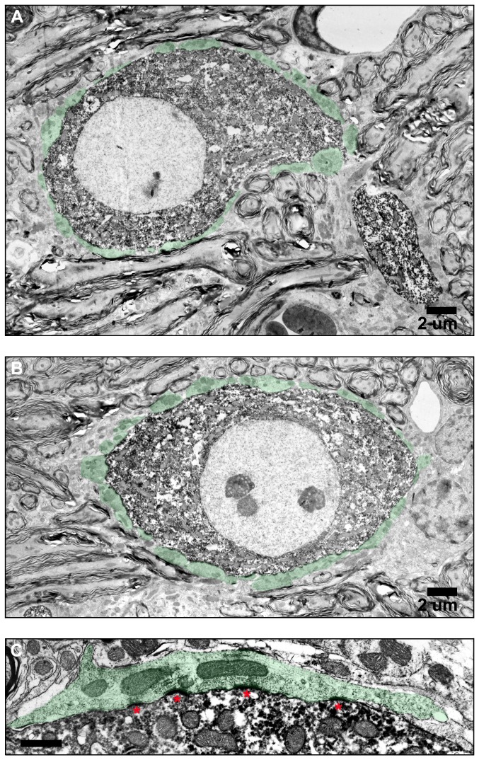
Figure 7. Electronmicrographs showing ultrastructural detail of labeled bushy cells.
Synaptic terminals contacting two labeled bushy cells in caudal AVCN are highlighted in green (A, B). (C) High magnification image of a primary auditory nerve terminal forming a synapse with a labeled bushy cell. PSDs are marked with asterisks.

